Research Article - International Research Journal of Plant Science ( 2022) Volume 13, Issue 3
Received: 25-Apr-2022, Manuscript No. IRJPS-22-61814; Editor assigned: 26-Apr-2022, Pre QC No. IRJPS-22-61814(PQ); Reviewed: 10-May-2022, QC No. IRJPS-22-61814; Revised: 13-May-2022, Manuscript No. IRJPS-22-61814(R); Published: 20-May-2022, DOI: http:/dx.doi.org/10.14303/irjps.2022.013
Withania somnifera (L.) Dunal (Solanaceae), commonly known as ashwagandha or Indian ginseng is a valuable medicinal plant and it is very potent, far and wide used herb in Ayurveda and other traditional systems of medicine in India. The qualitative and quantitative analysis has shown that methanol, ethanol and isopropanol has rendered a good yield of secondary metabolites. The silver nanoparticles used in W. somnifera AgNPs were characterized using UV-VIS spectrophotometry has shown two characteristic absorbance peaks value at 261.46 nm and 451.67 nm. The W. somnifera AgNPs has been detected with considerable antibacterial effect in comparison with the crude extract. The scanning electron microscopic (SEM) analysis also reveals the influence of W. somnifera AgNPs over the inhibition of biofilm formation in test pathogen Pseudomonas. The present study shows that isopropanolic root extracts of Withania somnifera has exhibited a substantial inhibitory effect towards the test pathogens used which may be due to the high degree of alkaloids retained in the extract that possess the inhibitory effect and renders bioactivity towards the pathogen. The antibacterial activity of isopropanolic root extracts of W. somnifera against the pathogens like E.coli, Klebsiella, Staphylococcus and Pseudomonas was detected and compared along with the standard antibiotics like ciprofloxacin and kanamycin. The maximum zone of inhibition and the activity index were observed in isopropanol extract against Pseudomonas (0.792). The MIC and MBC of crude isopropanol root extract of W. somnifera was determined to be 25 mg/ml and 6.25 mg/ml respectively against Pseudomonas. Similarly, the MIC and MBC of W. somnifera AgNPs extract tested against Pseudomonas was 25 mg/ml and 12.5 mg/ml respectively. The silver nanoparticles acts as an effective carrier of active ingredients of W. somnifera extract into the cells of pathogens which has probably inhibited its growth and thereby rendering an antibacterial activity to the W. somnifera AgNPs as effective as the crude solvent extracts.
Withania somnifera, Silver nanoparticles, SEM, UV-VIS spectrophotometry and antibacterial activity
Withania somnifera (L.) Dunal is also commonly known as Ashwagandha, Indian ginseng and Winter cherry is a small medium under a shrub belonging to the Solanaceae family. Nano encapsulation of the phytochemical was done in order to have increased bioavailability and increased water solubility, ensuring a better drug delivery system. Silver, a noble metal has been known to improve the immunity since ancient times. Silver nanoparticles have particular properties that may perhaps have a numerous applications in the fields of dentistry, clothing, catalysis, mirrors, optics, phytography, electronics and food industry (Bashir et al., 2013) Metallic silver in the form of silver nanoparticles has made a remarkable as potential antimicrobial agent (Datta et al., 2011). An extensive work has been done to develop new drug from natural products because of the resistance of microorganisms to the existing drug (Singh & Kumar, 2012). The current objective is focused on preparation of Withania somnifera silver nanoparticles (AgNPs) and characterization using UV-Visible spectrophotometer and scanning electron microscopy. The antibiotic effect of W. somnifera AgNPs was analyzed by using disc diffusion method and MIC/ MBC and the inhibition effect was compared with crude isoproponlic extract.
Plant material
Withania somnifera is a perennial shrub collected from the foot hills of Kolli hills of Namakkal District, Tamil Nadu, and India and maintained in the nursery was purchased and used for the study. The roots of W. somnifera were shadow dried and finely powdered to prepare the aqueous extract. The systematic identification of the plant was done by the Department of Botany, Vivekanandha College of Arts and Sciences for Women (Autonomous), Elayamapalayam, Tiruchengode.
Preparation of extracts
Roots of W. somnifera were crushed, powdered and extracted with various solvents like aqueous, ethanol, methanol and isopropanol for screening against the selected test pathogens for antibacterial activity. Fresh leaves of the sample were thoroughly washed 2-3 times with running water followed by sterilized distilled water. The washed leaves were dried under the shade at room temperature and then pulverized by mechanical grinding. The powdered plant (10 gms each) was extracted with 100 ml of aqueous, ethanol, methanol and isopropanol in a conical flask and kept at room temperature in a rotary shaker for 24 hours. After 24 hours, it was filtered through Whatman no.1 filter paper; and kept in hot air oven at 40°C for 24 hours to evaporate the solvents and dried. The dried leaf extract was dissolved in respective solvents to a final concentration of 1 mg/ ml. It was stored in the deep freezer for further analysis and screening (Jain & Agrawal, 2008).
Bacterial strain
Common pathogens like Bacillus sp., Staphylococcus sp., Escherichia coli, Klebsiella sp. and Pseudomonas sp. were used for determination of biofilm formation and to test the antibacterial effect of W. somnifera crude extract and its AgNPs.
Culture medium and inoculums preparation:
Nutrient broth / agar medium were used as a culture medium to maintain the microbial strains to perform the antimicrobial assay. Bacterial culture was inoculated into a nutrient broth (liquid medium) and incubated at 37°C for 24 hours and the culture density was checked for presence of approximately 10-5 CFU/ml Mac Farland’s standard by turbidity method.
Preparation of 1mM silver nitrate solution and synthesis of silver nanoparticles:
Silver nitrate (21.2 mg of AgNO3 powder in 125 ml of sterilized double distilled water) was prepared and stored in amber colored bottles in order to prevent auto oxidation (Jeyanthi et al., 2013). 1 mM Silver nitrate solution was added approximately one drop per second to about 0.002M Sodium borohydride (NaBH4) by constant stirring using magnetic beads. The mixing cause reduction of Ag ions and is clustered to form monodispersed or nanoparticles as a transparent sol in the aqueous medium. The addition of a few drops of 1.5 M Sodium chloride solution causes the suspension to turn darker yellow, then grey as the nanoparticles aggregate. The Ag solution becomes yellow because of absorption at 386nm (Mavani & Shah, 2013). Synthesis of Withania somnifera silver nanoparticles (WSAgNPs).
W. somnifera root extract and 1mM silver nitrate solution were taken in 1:4 ratio respectively and stirred continuously on the hot plate at 60ºC for 30 minutes until the color change was observed. This indicates the primary confirmation for the formation of Withania somnifera - silver nanoparticles (WS-AgNPs or As-Ag nanoparticles).
Characterization of silver nanoparticles using UVVisible spectra analysis
The reduction of pure Ag+ ions was monitored by measuring the UV-Vis spectrum of the reaction medium after 30 min using UV-Vis spectrophotometer (ELICO SL-159). A small aliquot of the sample was taken for UV-Vis spectrum analysis (200-500 nm). The maximum absorbance spectrum of As-Ag nanoparticles was usually observed at 455 nm.
SEM analysis of silver nanoparticles
Scanning Electron Microscopic (SEM) analysis was done using Hitachi S-4500 SEM machine. Thin films of synthesized and stabilized silver nanoparticles were prepared on a carbon coated copper grid by just dropping a very small amount of the sample on the grid and sample was analyzed for morphology and size of the silver nanoparticles.
Biofilm assay- Congo red method
The Brain heart infusion agar was prepared and sterilized at 121ºC for 15 minutes. Then the plates were inoculated with test organism and incubated at 37ºC for 24-48 hours aerobically. Black colonies with a dry crystalline consistency indicated biofilm production.
Tube Method
10ml of brain heart infusion broth with 1% glucose was inoculated with a loop full of test organism from overnight cultures on nutrient agar individually. The tubes were incubated at 37ºC for 24 hours. The culture was discarded and tubes were washed with phosphate buffer saline (pH- 7.3). The tubes were dried and stained with 0.1% crystal violet. Excess stain was washed with deionized water and the tubes were dried in an inverted position. In a positive biofilm formation, a visible stain film was seen lining the wall and the bottom of the tubes. Experiments were done thrice in triplicates and read or ranked as absent, weak, moderate and strong.
Screening of Antibacterial activity by disc diffusion method
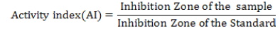
The antibacterial activity of the plant extract and WS-AgNPs extract was determined by disc diffusion method for initial screening. The 24 hours bacterial culture was diluted (10-5 CFU/ml) and 0.2 ml was spread over nutrient agar plates using sterile glass L-rod. Sterile Whatman No.1 filter paper discs (6.0 mm in diameter) were used for the experiment. The discs were impregnated with plant extracts and stored at 5°C prior to use. The extract impregnated discs were placed on an agar medium in the laminar air flow and incubated at 37°C for 24 hours. An antibacterial activity was determined by measuring the clear zones of inhibition formed around the discs. Standard antibiotic ciprofloxacin (5 mcg/disc) and kanamycin (30mcg/disc) (Span Diagnostics Ltd., Surat, India) was used as reference or positive control. The antibacterial activity of plant extracts was compared with standard antibiotic i.e. ciprofloxacin and kanamycin and their activity index were calculated.
Determination of the Minimum Inhibitory Concentration
The initial plant extract (1mg/ml) was serially diluted by transferring 5ml of the sterile plant extract (stock solution) into 5 ml of sterile nutrient broth (tubes) and nutrient agar (plates) to obtain the following dilutions: 100 μl, 50 μl, 25 μl, 12.5 μl, 6.25 μl and finally 3.125 μl. After obtaining the crude isopropanol extract and aqueous AgNPs, it was inoculated with 0.1 ml of a standardized bacterial cell suspension (approximately 105 CFU/ml) and incubated at 37ºC for 24 hrs and observed to change in turbidity and compared with the growth in control. The control was prepared as follows: nutrient broth, nutrient broth and plant extract, nutrient broth and test organisms. The lowest concentration of the extract that inhibited the growth of the test organism was taken as the MIC.
Determination of the Minimum Bacterial Concentration
The nutrient broth taken in equal volume of the various concentrations of isopropanol extract and aqueous AgNPs was mixed with micro-tubes to make up 0.5 ml of solution. Then 0.5 ml of McFarland standard of the organism suspension was added to each tube. The tube was incubated aerobically at 37˚C for 24 hrs. Two control tubes were maintained for each test batch. These include tube containing the growths without inoculums and the tube containing growth and inoculums. The MBC was determined by sub culturing the test dilution on nutrient agar and further incubated for 24 hrs. The highest dilution that yielded no single bacterial colony was considered as the Minimum Bacterial Concentration.
Synthesis and characterization of AgNPs
The roots of Withania somnifera were finely powdered and the crude solvent extracts were tested against the selected bacterial pathogens like Bacillus spp., Staphylococcus species, Escherichia coli, Klebsiella species and Pseudomonas spp. for the detection of their antibacterial activity. Similarly the aqueous root extract was used for the preparation of W. somnifera – silver nanoparticles and a comparison between crude extract and AgNPs was established based on their inhibitory activity (Figure 1).

Figure 1. Characterization of silver nanoparticles of crude extract of Withania somnifera (WS-102) by UV-Visible Spectrophotometry Analysis.
In the present analysis, these AgNPs were synthesized by using aqueous root extracts of Withania somnifera by incubating with silver nitrate. A color change of the medium from faint to dark brown color was developed by the addition of silver nitrate. The appearances of dark brown color indicated the presence of SNP.
Confirmation by UV-Visible spectrophotometer
The reduction of silver nitrate was monitored by measuring the UV-visible spectrum of the reaction medium, thereby confirming the synthesis of silver nano particles in the medium. UV-visible spectrum of colloidal solution of SNPs synthesized from a root extract of Withania somnifera have shown a characteristic absorbance peak at 261.46 nm and 451.67 nm and the broadening of the peak indicated that the particles are poly dispersed. The peak illustrates the presences of homogeneous distribution of hydrosol silver nano particles after stirring (Figure 2).

Figure 2. UV –Visible Spectrophotometry analysis of Crude extract for WS-AgNP.
Scanning Electron Microscopy (SEM)
Scanning Electron Microscopy was performed to confirm the development of silver nano structure. The control sample shows high density silver nano particles of Withania somnifera (Figure 3).

Figure 3. SEM analysis of Test pathogen and SNP of Withania somnifera (WS-102).
Evaluation of biofilm producers
The biofilm formation of the selected pathogens was assessed by the Congo red method and the tube adherence method. The black color colonies obtained by the Congo red method indicate, the formation of biofilms while the red color colonies refer to non-biofilm producers. In the present investigation culture plate streaked with Staphylococcus and Pseudomonas has exposed a very black color, thereby showing them as high producers, while E.coli and Bacillus was showing black coloration revealing them as producers and red color colony in the culture plate streaked with iKlebsiella shows that they are non producer of biofilm. In the tube adherence method visible stained film was seen lining the wall and bottom of the tube and indicates that the Staphylococcus and Pseudomonas as strong producers of biofilm, the E.coli and Bacillus are moderate producers of biofilm and Klebsiella was the weak producer of biofilm. The W. somnifera crude solvent extracts like methanol, ethanol and isopropanol were tested against the test pathogens and it was observed that the isopropanolic extract has shown a higher zone of inhibition and activity index than the other solvent. The isopropanolic extract was further subjected to MIC and MBC against the test pathogens and the results were compared with the activity of WS-SNPs (Figure 4) (Tables 1 and Table 2).

Figure 4. Evaluation of biofilm formation by Congo red methods and Tube adherence methods.
| Name of the pathogen | Biofilm phenotype | Slime production |
|---|---|---|
| Bacillus | Black | Producer |
| E.coli | Black | Producer |
| Klebsiella | Red | Non producer |
| Pseudomonas | Very black | Producer |
| Staphylococcus | Very black | Producer |
Table 1. Evaluation of biofilm formation by Congo red method.
| Name of the pathogen | Biofilm production | |
|---|---|---|
| Bacillus | ++ | Moderate |
| E.coli | ++ | Moderate |
| Klebsiella | + | Weak |
| Pseudomonas | +++ | Strong |
| Staphylococcus | +++ | Strong |
Table 2. Visual ranking of biofilm production of the Pathogen using tube adherence method (Note: +++ -Strong, ++-Moderate, +-Weaks).
Inhibitory effects of crude solvent extract of W. somnifera against pathogens by disc diffusion assay
The W. somnifera crude solvent extracts like methanol, ethanol and isopropanol were tested against the test pathogens and it was observed that the isopropanolic extract has shown a higher zone of inhibition and activity index than the other solvent. The isopropanolic extract was further subjected to MIC and MBC against the test pathogens and the results were compared with the activity of WS-SNPs.
Screening of Anti-microbial Activity by Disc Diffusion Assay from AgNP (WS-102)
The silver nano particles produced from the aqueous root extract of WS-102 was screened for its antimicrobial potential by disc diffusion assay and it was observed that the maximum activity was observed for Pseudomonas as represented by its activity index (AI: 1.286; ZI: 1.10 ± 0.063). It has shown at least a response against E. coli (AI: 0.357; ZI: 1.07 ± 0.127) and Klebsiella (AI: 0.394; ZI: 1.1 ± 0.124) (Figure 5) (Table 3).

Figure 5.Antimicrobial activities in AgNP of Withania somnifera (WS-102) using disc diffusion assay.
| Sl. No | Concentration | Plant parts | Test pathogens | ||||||||
|---|---|---|---|---|---|---|---|---|---|---|---|
| Solvent | E.coli | Klebsiella | Pseudomonas | Staphylococcus | |||||||
| ZI±SD | AI | ZI±SD | AI | ZI±SD | AI | AI | |||||
| ZI±SD | |||||||||||
| 1 | SNP | 1mg/ml | Root | 1.07± | 0.357 | 1.1± | 0.394 | 1.10± | 1.286 | 1.237± | 0.961 |
| 0.127 | 0.124 | 0.063 | 0.049 | ||||||||
| 2 | Standard | 0.529 | 0.467 | 1.342 | 1.124 | ||||||
| antibiotic | 1.12± | 1.10± | 1.24± | 1.286± | |||||||
| (ciprofloxacin | 0.186 | 0.135 | 0.129 | 0.056 | |||||||
| & kanamycin) | |||||||||||
Table 3. Screening of Anti-microbial Activity in AgNP of W.somnifera using Disc Diffusion assay (WS-102).
Determination of antibacterial activity of W. somnifera by Minimum Inhibitory Concentration (MIC) and Minimum Bactericidal Concentration (MBC)
Withania somnifera crude extract (WS-102) reveals a MIC value of 6.25mg/ml against Pseudomonas and Staphylococcus, whereas the MIC value against E.coli and Klebsiella recorded as 12.5 mg/ml respectively. However the MBC value was same against all the pathogen (12.5 mg/ml). Comparatively AgNP produced using aqueous root extract was tested against pathogens shows a similar result to that of crude extract. MIC & MBC of extract –SNP complex was in the range of 12.5 mg/ml to 25 mg/ml against test pathogens. The MIC value of WS-SNP against Pseudomonas was (25 mg/ml) and an MBC value was 12.5 mg/ml. The figure 6b illustrate the activity of SNP medicated Withania somnifera crude extract against the biofilm forming pathogen figure 6c Pseudomonas. The image reveals that deformation in the biofilm formation may be due the activity of SNP-extract complex (Figure 6 (a,b,c)) (Table 4).

Figure 6 (a, b, c): Determination of MIC and MBC for the WS-Silver nanoparticles Withania somnifera (WS-102) against the test pathogens (a. MIC analysis of WS-SNP; b. MBC analysis of WS-SNP; c. MIC analysis of WS-SNP).
| Sample | Solvent | Plant parts | Test pathogens | ||||||||
|---|---|---|---|---|---|---|---|---|---|---|---|
| E.coli | Klebsiella | Pseudomonas | Staphylococcus | ||||||||
| MIC | MBC | MIC | MBC | MIC | MBC | MIC | MBC | ||||
| WS- SNP | Isopropanol (1mg/ml) | Roots | |||||||||
| 12.5 | 12.5 | 12.5 | 25 | 25 | 12.5 | 12.5 | 25 | ||||
Table 4. Determination of MIC and MBC of the crude isopropanol extract and SNP of Withania somnifera against the test pathogens.
The formation of W. somnifera silver nanoparticles by using root aqueous extracts is confirmed by formation of dark brown color due to reduction of silver nitrate which indicates the presence of electron donor group in aqueous extract of fresh roots which itself act as a capping agent or stabilizing agent. The color change in the solution is due to the surface-Plasmon resonance (SPR) phenomena and the dark brown colour formation is because of excitation of surface plasmon vibrations in silver nanoparticles. Further characterization of silver nanoparticles was done by UVVIS spectrophotometry. UV-VIS spectrophotometry uses a principle called surface Plasmon resonance (SPR) for characterization which is described as resonant, collective oscillation of valance electron in a solid stimulated by incident light. The resonance condition is established by when frequency of light (ie. Photons) matches the natural frequency of surface electrons oscillating against the restoring force of positive nuclei. SPR in nanometer size structures is called localized surface plasmon resonance. The free electrons of SNPs gives rise to an SPR absorption bonds due to the combined vibration of electrons of metal nanoparticles in resonance with light waves. The UV-VIS spectrum of colloidal solutions of SNPs synthesized from root extract of W. somnifera has the characteristic absorbance peaks ranges from 400 nm and 470 nm and the broadening of the peak indicated that the particles are poly-dispersed. Sometimes weak absorption peak at shorter wavelengths due the presence of several organic compounds which are known to interact with silver ions were also observed. In our present study, the weak absorption peak was detected at 261 nm (Peak1) and the characteristic absorbance peak was perceived at around 451 nm.
This absorbance peaks coincide with the earlier reports pertaining to the characterization of silver nanoparticles (Li et al., 2007). Further, it is obvious that UV-VIS spectroscopy could be used to examine the size and shape of nanoparticles in aqueous solutions and also mentioned that the secondary metabolites present in the plant system may be responsible for the silver nitrate reduction and synthesis of silver nanoparticles (Rajesh et al., 2013). The test pathogens used for the antimicrobial activity were assessed for their biofilm producing capacity and it was observed that except Klebsiella all the pathogens including Bacillus, E. coli, Staphylococcus and Pseudomonas were found to be biofilm producers. The visual ranking done on the basis of Congo red and tube adherence method has clearly shown that Bacillus and E. coli is moderate producer of biofilms whereas the Staphylococcus and Pseudomonas were considered to be strong biofilm producers as they form very black colonies by Congo red method. Since Pseudomonas was found to be highly susceptible to isopropanolic crude root and leaf extracts of W. somnifera, it is apparently chosen as an ideal pathogen to analyze the effect of AgNP on their biofilm. It was obvious from the SEM analysis that AgNP has effect over the biofilm of Pseudomonas. A similar report by positively argues that the effect of silver nanoparticles on biofilm formations could be detected and studied using SEM (Patil et al., 2012). Biofilm formation is facilitated by certain quorum sensing compounds called auto-inducers (AI) which at threshold level protects the bacteria like Pseudomonas from antibiotics or bioactive compounds. In the present study, the bioactive secondary metabolites present in the solvent extract of W. somnifera may inhibit such quorum sensing auto-inducers thereby rupturing the biofilm and inhibiting their growth (Singariya et al., 2012). The bioactive compounds present in the root extracts of W. somnifera as well as its SNPs derived from aqueous root extract were found to be effective against Pseudomonas irrespective of the solvent used and shown a higher zone of inhibition comparatively equal to that of the commercial antibiotics.
But in the present study it was shown that isopropanol leaf extract of W. somnifera was effectual comparatively to ethanol and methanol extract while Klebsiella being more resistant to all the solvent extracts. It was also observed that low zone of inhibition was exhibited by the root extract of W. somnifera against Staphylococcus thereby displaying its resistance against the bioactive compounds. In a reported that the Methanolic root extracts of W. somnifera showed a greater zone of inhibition against Bacillus, E. coli and Pseudomonas which coincided with our current investigation (Rai et al., 2009). They also added that ethanolic root extracts show an equivalent or less active as compared to commercial antibiotics. But their result diverges from ours with respect to the activity of Methanolic root extract which was found to be active against Klebsiella. The isopropanolic root extracts of W. somnifera was found to be effective against the pathogens due to the reason that it possesses the bioactive components like alkaloid, flavonoids, tannins and phytosteroids which were already reported to have inhibitory activity against the pathogens. Further, these solvent extracts (ethanol, methanol and isopropanol) possess more stable steroidal lactones like withanolides when compared to the other solvent extracts making them a suitable preference for the assay. It was reported that the presence of flavonoids, steroids, alkaloids, saponins and tannins in the Methanolic extracts of W. somnifera and assessed the antibacterial activity which has revealed a moderate bioactivity against various pathogens (Santhi & Swaminathan, 2011). They also further evaluated the three different flavonoids like 7, 3', 4'-trihydroxy flavone- 3-O-rhmnosyl, Quercetin -3-O-galactosyl and 5, 7, 4`- triahydroxy-methyl-3-O-galactosyl flavonols and observed very low, moderate and high inhibition against most of the pathogens tested. This work supports our investigation by relating the inhibitory activity to the concentration of the bioactive components available in extract. The low molecular weight and moderately polar substances due to their wide range of solubility could be easily extracted using methanol as solvent (Terasa et al., 2000). However, current investigation reports that isopropanolic extracts of roots of W. somnifera also exhibit a substantial inhibitory effect towards the test pathogens used. This may be due to the high degree of alkaloids retained in the extract that possess the inhibitory effect and renders bioactivity towards the pathogen. Since the root extracts of W. somnifera has revealed a higher bioactivity when compared to leaf extract by disc diffusion assay, the isopropanolic root extract of W. somnifera was resolved using MIC/ MBC tests to establish its bacteriostatic effect. Similarly the AgNP of WS- 102 was also subjected to MIC and MBC and the results were almost in concurrence with the crude extract except for the Staphylococcus and Pseudomonas where the crude extract was more effective than that of the AgNP extract. The MBC values were almost coinciding for both the extracts towards the test pathogens used (Subbaiah & Savithramma, 2013). The results of many reports have suggested the use of MIC and MBC to determine the bacteriostatic effects of W. somnifera using broth dilution technique (Wiley et al., 2006).
Further, it is concluded that the silver nanoparticles acts as an effective carrier of active ingredients of W. somnifera into the cells of pathogens which has probably inhibited its growth and thereby rendering an antibacterial activity to the AgNP as effective as the crude solvent extracts.
We would like to thank Prof. Dr. M. Karunanithi, our Honorable Chairman, Vivekanandha Educational Institutions for providing us the infrastructure and facilities to complete our current research work.
Bashir HS, Mohammed AM, Magsoud AS, Shaoub AM (2013). Isolation of three flavonoids from Withania somnifera leaves (Solanaceae) and their antimicrobial activities.J Forest Products Industries.2: 39.
Datta S, Kumar Pal NK, Nandy AK (2011). Inhibition of the emergence of multi drug resistant Staphylococcus aureus by Withania somnifera root extracts. Asian Pac J trop Med. 4: 917-20.
Indexed at, Google Scholar, Cross Ref
Singh G, Kumar P (2012). Antibacterial Potential of Alkaloids of Withania somnifera L. and Euphorbia hirta L. Int J Pharmacy Pharm Sci. 4: 78-81.
Jain PK, Agrawal RK (2008). Antioxidant and free radical scavenging properties of developed mono and poly herbal formulations. Asian J exp Sci. 22: 213-20.
Jeyanthi T, Subramanian P, Kumaravel P (2013). A comparative analysis of antibacterial activity of Withania somnifera root extract with commercial antibiotics. Asian J Pharm Res. 3: 98-102.
Mavani K, Shah M (2013). Synthesis of silver nanoparticles by using sodium borohydride as a reducing agent. Int J Eng Res. 2: 1-5.
Li S, Shen Y, Xie A, Yu X, Qui L et al., (2007). Green synthesis of silver nanoparticles using Capsicum annum L. extract. Green Chem. 9: 852-858.
Indexed at, Google Scholar, Cross Ref
Rajesh P, Swati W, Sandesh M, Sangita J, Kulkarni S (2013). Green synthesis of silver nanoparticles by Withania somnifera and evaluation of its antimicrobial potential. J Empir Biol. 1: 38-48.
Indexed at, Google Scholar, Cross Ref
Patil RS, Kokate MR, Kolekar SS (2012). Bio inspired synthesis of highly stabilized silver nanoparticles using Ocimum tenuiflorum leaf extract and their antibacterial activity. Spectrochim Acta A Mol Biomol Spectrosc. 91: 234-238.
Indexed at, Google Scholar, Cross Ref
Singariya P, Mourya KK, Kumar P (2012). Antimicrobial activity of the crude extracts of Withania somnifera and Cenchrus setigerus in-vitro. J Pharmacogn. 4: 60-65.
Indexed at, Google Scholar, Cross Ref
Rai M, Yadav A, Gade A (2009). Silver nanoparticles as a new generation of antimicrobials. Bio technol Adv. 27: 76-83.
Indexed at, Google Scholar, Cross Ref
Santhi M, Swaminathan C (2011). Evaluation of antibacterial activity and phytochemical analysis of leaves of Withania somnifera (L.) Dunal. Int J Curr Res. 3: 10-12.
Terasa R, De Kievit, Barbara H, Iglewsk (2000). Bacterial quorum sensing in pathogenic relationship. Infect Immun. 68: 4839-49.
Subbaiah KPV, Savithramma N (2013). Antimicrobial efficacy of silver nanoparticles synthesized from Withania somnifera-an important ethnomedicinal herb of Kurnool district, Andhra Pradesh, India. Int J Pharm Sci Rev. 22: 216-222.
Wiley BJ, IM SH, Li ZY, Mclellan J, Siekkinen A et al., (2006). Maneuvering the Surface Plasmon Resonance of Silver Nanostructures through Shape-Controlled Synthesis. J Phys Chem.110: 15666-75.
Indexed at, Google Scholar, Cross Ref
Citation: Logeswari N & Prabhakaran S.G (2022). Characterization of Withania somnifera (L.) Dunal silver nanoparticles and its antimicrobial effect on biofilm forming pathogenic microbes. IRJPS. 13: 013.