Journal of Medicine and Medical Sciences is making SARS-CoV-2 and COVID-19 research free*
The Journal of Medicine and Medical Sciences (JMMS) (ISSN-2141-9477) is an international multidisciplinary peer-reviewed, Open Access Journal with reputable academics.The journal is indexed in and Google Scholar with a Journal Impact Factor of 0.45*. It promotes excellence in current research works for advancing knowledge on Medicine and Medical Sciences. Journal of Medicine and Medical Sciences is a leading international journal devoted to publication of original research and reviews covering applied, methodological and theoretical issues which emphasis on studies using multidisciplinary or integrative approaches.
This Journal aims to establish channels of communication between academic and research scientific experts by making accessible to the health community, policy makers and to improve medical research worldwide. The journal is dedicated to increasing the depth of medicine and medical sciences across disciplines with the coverage of all areas basic, clinical, experimental, preventive, environmental and social medicine.
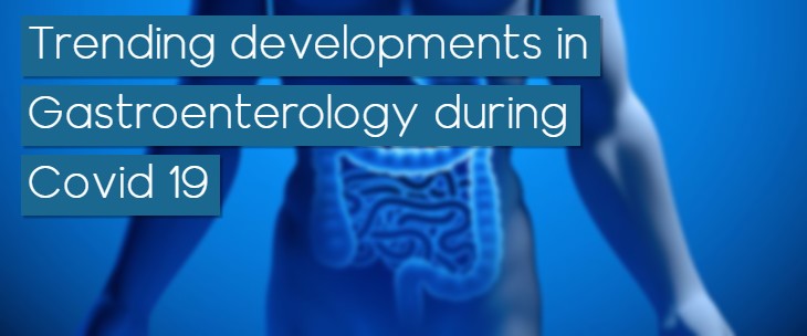
Scope: Medical Microbiology, Medical Biochemistry, Allergy and Immunology, Cardiovascular Medicine, Endocrinology, Diabetes and Metabolism, Gastroenterology, General Medicine and Primary Care, Gerontology, Haematology/Oncology, Infectious Diseases and Travel Clinic, Nephrology, Pulmonary, Critical Care and Sleep Medicine, Rheumatology, Anaesthesia, Anatomy, Chemical Pathology, Community Medicine, Dermatology, Haematology and Immunology, Medicine, Anatomy, Obstetrics and Gynaecology, Ophthalmology, Otorhinolaryngology, Paediatrics, Paediatric Surgery, Pharmacology/Therapeutics, Physiology, Psychological Medicine, Radiation Medicine, Surgery, Diabetes, Thyroid infections.
Policy Regarding the NIH Mandate: Journal of Medicine and Medical Sciences will support authors by posting the published version of articles by NIH grant-holders and European or UK-based biomedical or life sciences grant holders to PubMed Central immediately after publication.
Timeline:
Submit manuscript at Editorial Tracking or send as an e-mail attachment to the Editorial Office at manuscripts@interesjournals.org
Fast Editorial Execution and Review Process (FEE-Review Process):
Journal of Medicine and Medical Sciences is participating in the Fast Editorial Execution and Review Process (FEE-Review Process) with an additional prepayment of $99 apart from the regular article processing fee. Fast Editorial Execution and Review Process is a special service for the article that enables it to get a faster response in the pre-review stage from the handling editor as well as a review from the reviewer. An author can get a faster response of pre-review maximum in 3 days since submission, and a review process by the reviewer maximum in 5 days, followed by revision/publication in 2 days. If the article gets notified for revision by the handling editor, then it will take another 5 days for external review by the previous reviewer or alternative reviewer.
Acceptance of manuscripts is driven entirely by handling editorial team considerations and independent peer-review, ensuring the highest standards are maintained no matter the route to regular peer-reviewed publication or a fast editorial review process. The handling editor and the article contributor are responsible for adhering to scientific standards. The article FEE-Review process of $99 will not be refunded even if the article is rejected or withdrawn for publication.
The corresponding author or institution/organization is responsible for making the manuscript FEE-Review Process payment. The additional FEE-Review Process payment covers the fast review processing and quick editorial decisions, and regular article publication covers the preparation in various formats for online publication, securing full-text inclusion in a number of permanent archives like HTML, XML, and PDF, and feeding to different indexing agencies.
Articles published in Journal of Medicine and Medical Sciences have been cited by esteemed scholars and scientists all around the world. Journal of Medicine and Medical Sciences has got h-index 29, which means every article in Journal of Medicine and Medical Sciences has got 29 average citations.